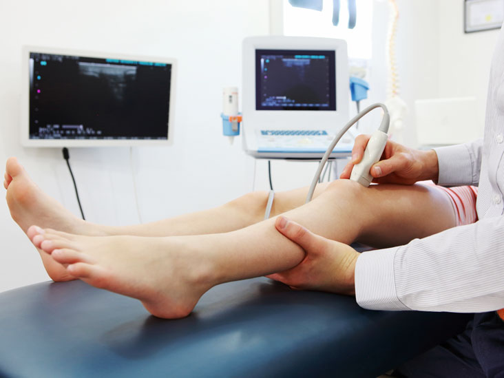
While the patient’s history and physical examination are the initial steps of making a medical diagnosis, the ability to peer inside the body can be a powerful tool. Ultrasound is an imaging technique that provides that ability to medical practitioners.
Ultrasonography uses high-frequency sound (ultrasound) waves to produce images of internal organs and other tissues. A device called a transducer converts electrical current into sound waves, which are sent into the body’s tissues. Sound waves bounce off structures in the body and are reflected to the transducer, which converts the waves into electrical signals. A computer converts the pattern of electrical signals into an image, which is displayed on a monitor and recorded on film, on videotape, or as a digital computer image. The technology is especially accurate at seeing the interface between solid and fluid filled spaces.
Ultrasonography is painless, relatively inexpensive, and considered very safe, even during pregnancy.
Usually, a radiologist will oversee the ultrasound test and report on the results, but other types of physicians may also use ultrasound as a diagnostic tool. For example, obstetricians use ultrasound to assess the fetus during pregnancy. Surgeons and emergency physicians use ultrasound at the bedside to assess abdominal pain or other concerns.

A transducer, or probe, is used to project and receive the sound waves and their echoes. A gel is wiped onto the patient’s skin so that the sound waves are not distorted as they cross through the skin. This may take a fair amount of time and require the probe to be repositioned and pointed in different directions. As well, the technician may need to vary the amount of pressure used to push the probe into the skin. The goal will be to "paint" a shadow picture of the inner organ that the health care practitioner has asked to be visualized.
Conventional ultrasound displays the images in thin, flat sections of the body. Advancements in ultrasound technology include three-dimensional (3D) ultrasound that formats the sound wave data into 3D images.
If the abdomen is being examined, you may be asked to refrain from eating and drinking for several hours before the test. Usually, the examiner places thick gel on the skin over the area to be examined to ensure good sound transmission. A handheld transducer is placed on the skin and moved over the area to be evaluated.
To evaluate some body parts, the examiner inserts the transducer into the body – for example, into the vagina to better image the uterus and ovaries or into the anus to image the prostate gland. To evaluate the heart, the examiner sometimes attaches the transducer to a viewing tube called an endoscope and passes it down the throat into the esophagus. This procedure is called transesophageal echocardiography.
After the test, most people can resume their usual activities immediately.
Ultrasound images are acquired rapidly enough to show the motion of organs and structures in the body in real time. For example, the motion of the beating heart can be seen, even in a fetus. Ultrasonography is effectively used to check for growths and foreign objects that are close to the body’s surface, such as those in the thyroid gland, breasts, testes, and limbs, as well as some lymph nodes.

Ultrasonography may be used to image internal organs in the abdomen, pelvis, and chest. However, because sound waves are blocked by gas (for example, in the lungs or intestine) and by bone, ultrasonography of internal organs requires special skills. People who have been specifically trained to do ultrasound examinations are called sonographers.
Ultrasonography is commonly used to evaluate the following:
- Heart: For example, to detect abnormalities in the way the heart beats, structural abnormalities such as defective heart valves, and abnormal enlargement of the heart’s chambers or walls.
- Blood vessels: For example, to detect dilated and narrowed blood vessels.
- Gallbladder and biliary tract: For example, to detect gallstones and blockages in the bile ducts
- Liver, spleen, and pancreas: For example, to detect tumors and other disorders
- Urinary tract: For example, to distinguish benign cysts from solid masses in the kidneys or to detect blockages such as stones or other structural abnormalities in the kidneys, ureters, or bladder
- Female reproductive organs: For example, to detect tumors and inflammation in the ovaries, fallopian tubes, or uterus
- Pregnancy: For example, to evaluate the growth and development of the fetus and to detect abnormalities of the placenta
References:


